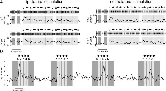
Figure 9.
Sensitivity to small moving objects. A, Raster plots and Gauss-filtered spike trains of the first-trial responses of a TB1 neuron to small black translating squares (8 × 8° in screen center) against a bright background. Dark gray (light gray; blank) areas represent time intervals when a stationary (moving; none) square was displayed. The first two dark areas represent time intervals during which the square was shown in the middle of the screen. This corresponds to an azimuth of 90° and an elevation of 0° when defining a position in front of the locust as being at an azimuth of 0° and an elevation of 0°. Labels above the raster plots indicate the extreme positions of the square (d, dorsal, azimuth 0°, elevation 47°; v, ventral, azimuth 0°, elevation −47°; a, anterior, azimuth 39°, elevation 0°; p, posterior, azimuth −39°, elevation 0°). Arrows indicate moving direction of the square. Note that in the top the square started moving ventrally while it started dorsally in the bottom. In each trial the neuron was excited in correlation with the stimulus only during the first vertical movements (v→d; d→v). In the contralateral trials there may also be an excitation during the first horizontal movement (a→p). B, Gaussian-filtered spike train of a CU4 neuron that responded in a directionally sensitive manner to a small, black translating square (8 × 8° in screen center) against a bright background. The eye contralateral to the cell body position was stimulated. Dark gray (light gray; blank) areas represent time intervals when a stationary (moving; none) square was presented. The square moved in a dorsoventral direction. h, v, d represent horizontal (0°), ventral (−39°), and dorsal (39°) elevations of the square. Arrows indicate moving direction. The neuron was excited when the square moved ventrally starting from a horizontal position.

