
Figure 3.
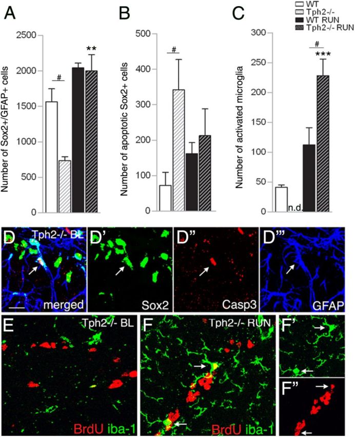
Sox2-expressing cells are highly affected in Tph2−/− mice. A, At P42, the number of overall Sox2/GFAP-expressing cells in Tph2−/− mice was significantly lower at baseline but doubled following exercise. B, At baseline, a significantly higher number of Sox2-expressing apoptotic cells (coexpression of Sox2/Caspase3), and increased microgliosis following running (C) was found in Tph2−/− mice. D, Confocal microscopy images of Sox2/Caspase3/GFAP coexpression (arrow), and (E) BrdU/iba-1 in Tph2−/− mice at P42 baseline levels (BL). F, Increased microgliosis in Tph2−/− mice following exercise (arrows indicate coexpression of BrdU/iba-1). Notably, the number of total iba-1-positive microglia is generally increased following exercise independently of genotype, and less frequent in sedentary mice. n.d., not detectable. Scale bar, 25 μm. **p < 0.01, ***p < 0.001 indicate statistical significance relatively to sedentary controls of the same genotype, and #p < 0.05, between WT and Tph2−/− for same condition (SEM).