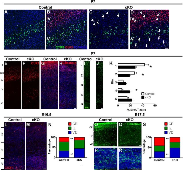
Figure 8.
Delayed migration and ectopic localization of late-born neurons in Mpp3 cKO cortex. A–C, In both control (A) and Mpp3 cKO (C) cortex, CTIP2 neurons are localized in lower layers. However, in Mpp3 cKO cortex, there are some ectopically localized CTIP2 neurons in upper layers (arrowheads in C). C, D, Removal of MPP3 results in stratification defects with ectopic CUX1 (arrows in D) and CTIP2 (arrowheads in D) neurons. E, Distribution of CUX1+ cells in control P7 cortex showed that the majority of CUX1+ cells reside in layer II–IV with some ectopic CUX1+ cells in layer V–VI. F, Also in Mpp3 cKO cortex, the majority of CUX1+ cells occupied layers II–IV, but there is an increase in number of ectopically localized CUX1+ cells in deeper layers. G, H, Distribution of Calretinin interneurons in control (G) and Mpp3 cKO (H) cortex shows an increased number of interneurons in layer VI. I, J, BrdU injection at E16.5 and analysis of distribution of BrdU+ cells in P7 control (I) and Mpp3 cKO (J) cortex. K, Quantification of the distribution of the BrdU+ cells shows that, in Mpp3 cKO cortex, fewer cells occupied superficial layers and there are an increased number of BrdU+ cells in deeper layers. L–N, Analyzing the distribution of CUX1+ cells between E16.5 control (L) and Mpp3 cKO (M) cortex showed that the total number of CUX1+ cells was not different. However, the number of CUX1+ cells still residing in the ventricular zone was increased, whereas the number of CUX1+ cells occupying the cortical plate was decreased (N). O–S, Distribution of Calretinin+ interneurons in E17.5 control (O, P) and Mpp3 cKO (Q, R) cortex. P and R are magnifications of Calretinin interneurons in the ventricular zone from O and Q, respectively. There is no difference in the number of Calretinin+ interneurons between control and Mpp3 cKO cortex, but the distribution is altered during removal of Mpp3, with a lower percentage of Calretinin+ interneurons in the cortical plate, and an increased percentage in the ventricular zone (S). VZ, Ventricular zone; IZ, intermediate zone; CP, cortical plate. *p < 0.05, **p < 0.001. Scale bars, 50 μm.

