Figure 4.
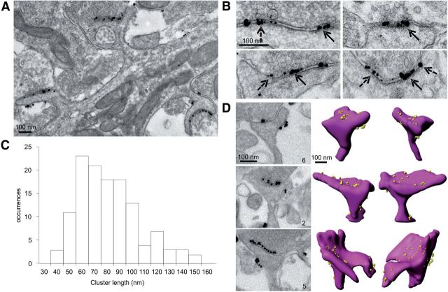
Nanodomain size and per-spine abundance revealed by pre-embedding immunogold EM. A, Low-magnification view of a portion of dendrite containing three synaptic spines that was live-labeled for GluA2 before fixation and then labeled with secondary fab′ fragment conjugated to a 1.4 nm gold particle. Silver-intensified label particles are highly concentrated at PSD-membrane in spines (7.23 particles/μm). Label particles are also present on extrasynaptic spine membrane (2.12 particles/μm) and at a relatively low density on the dendritic plasma membrane (1.24 particles/μm). B, High-magnification views of individual spine synapses show that label particles are present in nanodomains (solid arrows) or as single-label particles (dashed arrows). C, Histogram of the clusters of immunogold particles size in 2D EM sections showing that the average length of clusters of two or more label particles fits a single Gaussian with a mean of 77.48 ± 23.98. D, Images of three spines that were serial sectioned and used for 3D reconstructions (right) and count the number of nanodomains per spine. The number in lower right corner of each EM image indicates the number of nanodomains counted on each spine.

