
Figure 5.
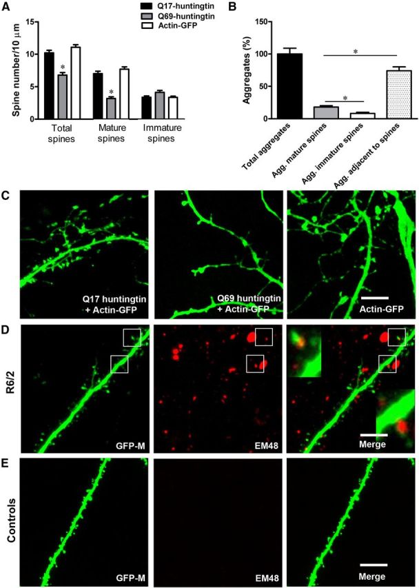
In vitro mutant huntingtin leads to loss of spines, especially of mature-type spines, and in vivo aggregates of mutant huntingtin localize in dendritic spines. A, Mean spine number/10 μm counted on ∼37–51 high-magnification dendritic segments of cortical/hippocampal neurons (13 neurons, 3–5 per genotype 4 × technical replicates) overexpressing short Q17 huntingtin, long Q69 huntingtin, or actin-GFP only. In neurons overexpressing Q69 huntingtin, we observed significant loss, particularly of mature-type spines, compared with neurons overexpressing Q17 huntingtin and actin-GFP only. B, Quantification of aggregates associated with dendritic spines in the R6/2 mice. More huntingtin aggregates were present within mature (mushroom-shaped) spines compared with smaller spines (filopodia-like or stubby spines; Agg, aggregates). However, there are more huntingtin aggregates adjacent to spines than within mature or immature spines. C, Z-projected stack representing total spines on cortical/hippocampal dendrite overexpressing Q17 + actin-GFP, Q69 + actin-GFP, and actin-GFP only. Notice that Q69 huntingtin overexpressing neurons (middle) have fewer spines compared with neurons expressing Q17 huntingtin + actin-GFP (left) and actin-GFP only (right). Also notice the presence of fewer mature spines on neurons overexpressing Q69 huntingtin. D, GFP expressing apical dendrite from double transgenic Thy1-GFP-M × R6/2 mice. In red is mutant huntingtin staining with anti-human huntingtin antibody EM48. Insets in the merged image represent boxed regions. Note the presence of mutant huntingtin aggregates in dendritic spines of R6/2 mice. E, GFP-expressing apical dendrite from the wild-type Thy1-GFP-M mice. No huntingtin aggregates are present in the wild-type mice. In this study, we did not observe any significant difference in shaft diameter between R6/2 and wild-type mice as evident in the figures above. Scale bar, 5 μm.