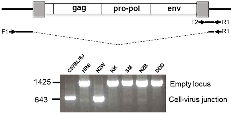
Fig 3. Detection of a representative ERV, Pmv7, in 7 inbred strains.
At the top is a diagram of the provirus and cellular flanks with arrows showing positions of 3 primers and the expected products for mice with and without the Pmv7 insertion. At the bottom are PCR test results for 7 strains using the three primers.

