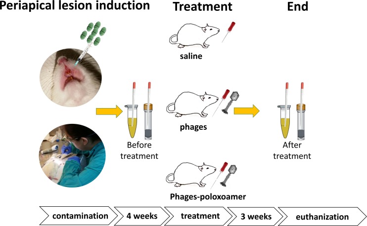
Fig 1. Periapical lesions were induced in a rat model.
Dental pulp of the maxillary incisor teeth of male Wistar rats were exposed and infected with E. faecalis (VRE ATCC 700802). Standard root canal treatment was conducted using instrumentation and one of the following treatments: group A: saline irrigation; group B: EFDG1/EFLK1 phage cocktail irrigation (109 PFU/mL); group C: EFDG1/EFLK1 poloxamer-phage formulation (109 PFU/mL).

