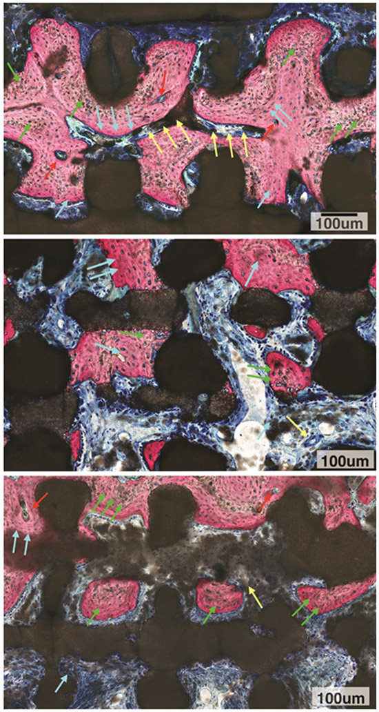
Figure 5A:
High magnification microscopy evaluation of DIPY group depicted active bone remodeling units (red arrows) leading to replacement of woven bone (green arrows) by lamellar bone (blue arrows) surrounding vascular structures (yellow arrows). Also, an irregular scaffold material surface was observed throughout the scaffold structure irrespective of what tissue type was in contact with the bioactive ceramic structure (dark stained structure).
5B: High magnification microscopy evaluation of COLL group depicted woven bone (green arrows) and replacement with lamellar bone (blue arrows). Also, irregular scaffold material surface was observed throughout the scaffold structure irrespective of what tissue type was in contact with the bioactive ceramic structure (dark stained structure).
5C: High magnification microscopy evaluation of control group depicted woven bone (green arrows) with some replacement by lamellar bone (blue arrows) in the presence of an active bone remodeling unit (red arrows), Vascular structure is depicted as well (yellow arrow). An irregular scaffold material surface was observed throughout the scaffold structure irrespective of what tissue type was in contact with the bioactive ceramic structure (dark stained structure).

