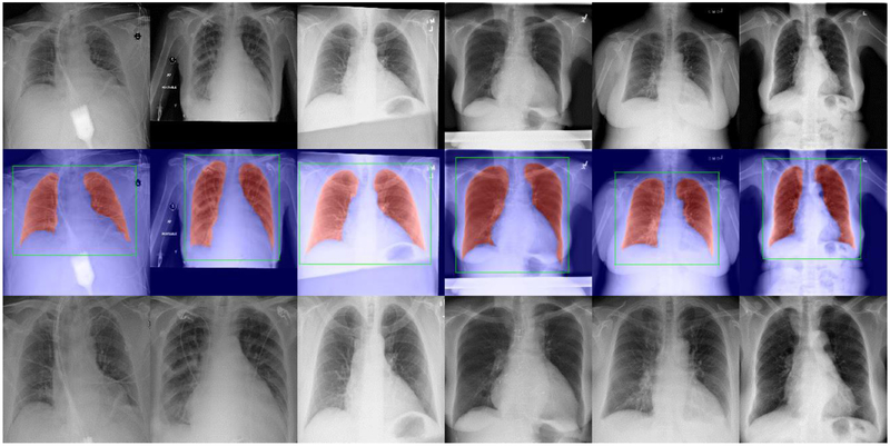
Fig. 6.
Examples demonstrating the lung segmentation and the identified lung region images. Top: some original CXR images in the Chest X-ray 14 Dataset. Middle: the lung segmentation results (in red) and the identified bounding boxes of the lung regions (in green). Bottom: the identified lung region images.

