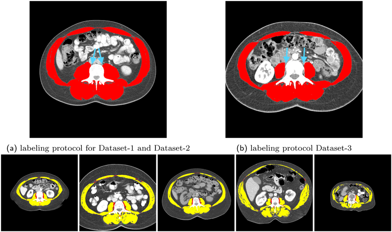
Figure 9:
The top row is the illustration of the differences in the manual labeling protocol of Dataset-3 and the other two datasets. The bottom row is the overlay of automated segmentation of samples images from Dataset-3 with the model trained on Dataset-2 and the ground truth segmentation for these images demonstrating the protocol-based discrepancy lowering the segmentation performance.

