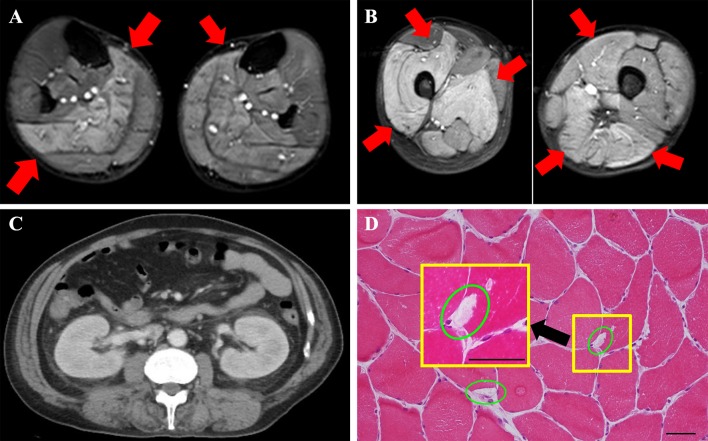
Fig. 1.
Findings upon radiological imaging and muscle biopsy. a Left upper extremity findings on MRI Short-Tau Inversion Recovery (STIR). MRI STIR imaging showed diffuse high signals in the bilateral thighs, which suggested myolysis (red arrow). b Left lower extremity findings on MRI STIR. MRI STIR imaging showed diffuse high signals in the bilateral thighs, which suggested myolysis (red arrow). c Computed tomography (CT) findings in the kidney. Swelling of the kidney was observed, but atrophy was not observed. No post-renal failure findings were observed. d Muscle biopsy tissue (hematoxylin and eosin stain). The scale bars are 100 µm (larger square) and 20 µm (smaller square). Muscle biopsy tissue showed almost normal findings. Vacuolated muscle fibers, a finding that suggests rhabdomyolysis, were present, although they were rare

