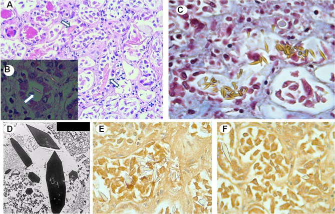
Fig. 2.
a Renal tissue shows intratubular large needle-shaped or rhomboid-like crystals (arrows) (H–E staining × 2000). b Intratubular crystals (arrow) show negative birefringence under polarized light (× 4000). c Crystal stain strongly yellow with the Masson’s trichrome stain. d Transmission electron microscopy microphotograph showing geometrically shaped crystals without periodic organization within the tubular lumen (× 25,000). Immunohistochemistry shows negative reactivity of crystals for kappa (e) and lambda (f) light chains (× 4000)

