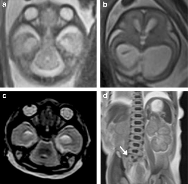
Fig. 2.
VACTERL spectrum anomalies in a 22-week gestation fetus. a, b Axial T2-weighted iuMR images at two different levels demonstrate a typical ‘ball’-shaped appearance of rhombencephalosynapsis (RES) and also lateral and third ventricular dilatation suggestive of aqueductal stenosis. c The T2-weighted PMMR image also shows a deficient cerebellar vermis with hemispheric fusion, although this was not appreciated. d Coronal T2-weighted PMMR of the body did however demonstrate a vertebral segmental anomaly (white arrow) that was not seen on iuMR

