Fig. 4.
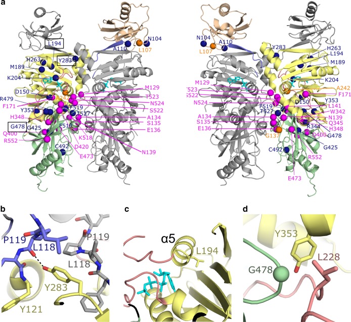
Location of variants associated with arhinia and FSHD2. a Cartoon diagram colored as in Fig. 1 with right panel ~180° rotation with respect to the left. Variants associated with arhinia, FSHD2, or with both diseases are colored magenta, dark blue, or orange, respectively. Some variants are only visible/labeled on one panel. b, c, d Close-up views of three regions in SMCHD1 where FSHD2 disease variants Y283C, L194F, and G478E (respectively) are located. The labels for these variants are boxed with black line on left figure of panel a. Black dashed lines indicate hydrogen bonds

