Figure 2.
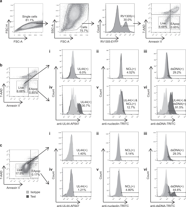
Expression of UL44 and interacting host antigens on ARPE-19 cell surface. (a) Gating strategy for infected APRE-19 cells. ARPE-19 cells were infected with RV1305, a strain of HCMV that expresses an EYFP fusion protein. Cells were stained with 7-AAD and annexin V before flow cytometric analysis. Live cells stain 7-AAD(−), annexin V(−) while early apoptotic (EApop) cells stain 7-AAD(−), annexin V(+). The gating strategy for uninfected cells was the same except that the cells were not gated based on EYFP expression before live/dead gating. (b) Flow cytometric analysis of RV1305-infected ARPE-19 cells post surface staining. Relative to isotype control antibody-stained cells, UL44 was observed on 30.7% of early apoptotic cells (iv), but not on live cells (i). Gating on UL44(+) cells revealed that 12.7% and 91.5% of the apoptotic cells displayed nucleolin (NCL) (v) and dsDNA (vi) respectively. In contrast, only 4.52% and 29.2% of the live cells displayed NCL (ii) and dsDNA (iii) respectively. (c) Flow cytometric analysis of uninfected ARPE-19 cells was performed to check for the specificity of 3A11 (i and iv) and surface expression of nucleolin (ii and v) and dsDNA (iii and vi). Nucleolin expression was not observed on uninfected cells. Relative to the isotype control, 29.3% of live (iii) and 44.6% of early apoptotic (vi) ARPE-19 cells stained positive for dsDNA.

