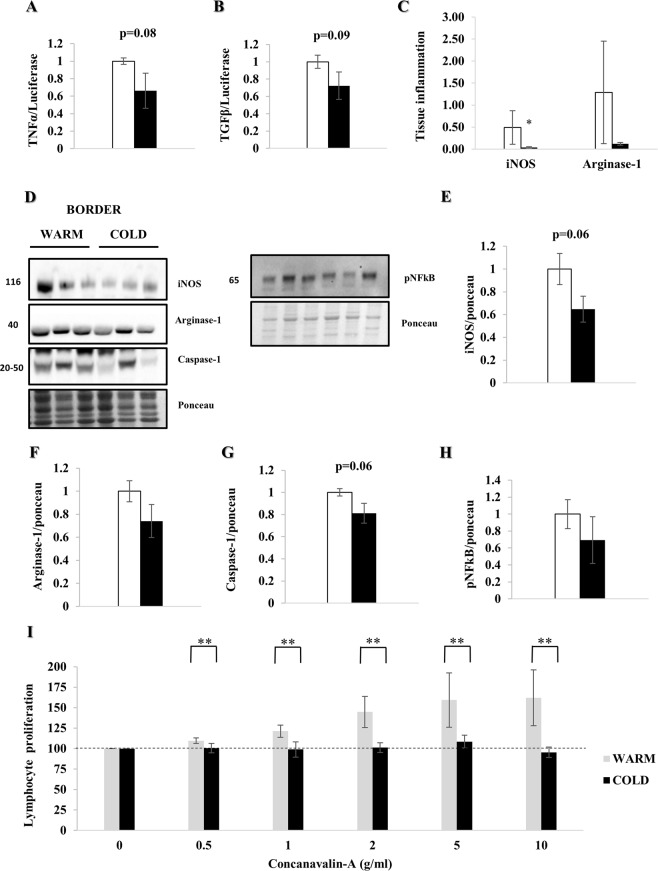
Figure 8.
Myocardial hypothermia decreases local and systemic inflammation. Myocardial border zone tissue of female farm pigs (n = 3 per group) was harvested one week post MI. (A,B) Quantification of TNFα and TGFβ mRNA expression respectively. (C) Quantification of local tissue inflammation via iNOS and Arginase-1 staining (representative images: Supplemental Fig. 2). (D) WB analysis with protein quantification (E–G) of iNOS, Arginase-1 and Caspase-1 respectively in whole lysate and (H) pNFκB in the nuclear fraction. (I) Quantification of lymphocyte proliferation with Concanavalin-A treatment on fresh splenic tissue, harvested from the same pigs. The data represents the mean ± SEM with *p < 0.05, **p < 0.01, comparing cold (black) vs. warm (white). We normalized each protein to the entire corresponding lane on the ponceau-stained membrane, representing the total protein amount. Due to space limitation, the blots are cropped from various gels, please see the supplemental data for full-length gels/blots.

