Figure 1.
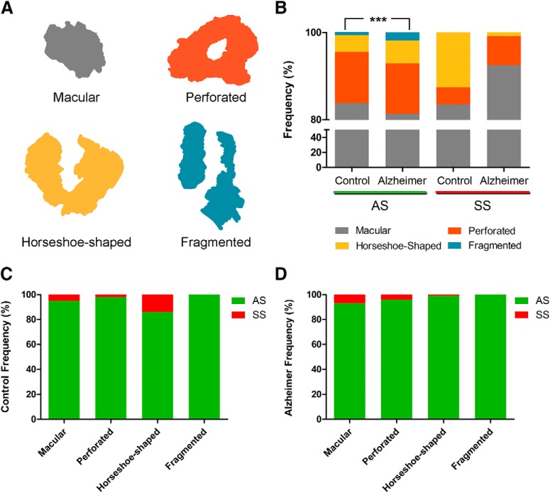
A, Schematic representation of the shape of the synaptic junctions: macular synapses with continuous disk-shaped PSD; perforated synapses with holes in the PSD; horseshoe-shaped with tortuous horseshoe-shaped perimeter with an indentation; and fragmented synapses with two PSDs with no connections between them. B, Proportion of macular, perforated, horseshoe-shaped, and segmented AS and SS in control cases and AD patients. In AD patients, fragmented synapses were significantly more frequent than in control cases (χ2, p < 0.001). C, Proportion of AS and SS belonging to each morphological category in control cases. The horseshoe-shaped synapses were significantly more frequent among SS than AS (χ2, p < 0.0001). D, Proportion of AS and SS belonging to each morphological category in AD cases. *** p < 0.001.

