Figure 4.
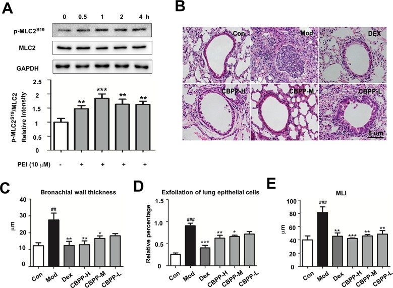
PEI and CBPP increased phosphorylation of MLC2 and improved lung function. (A) HBSMC cells were stimulated by 10 µM PEI, phosphorylation of MLC2 was measured by Western blot, and the relative intensity data of P-MLC2S19 to GAPDH are represented by the mean ± SD of three groups, **p < 0.01 and ***p < 0.001. (B) HE staining images (400-fold) of bronchus, the statistics of bronchial wall thickness (C), exfoliation of lung epithelial cells (D), and MLI (E) in bronchus pathological section. *p < 0.05, **p < 0.01, and ***p < 0.001 compared to the Mod. ## p < 0.01 and ### p < 0.001, compared to the Con (n = 6).

