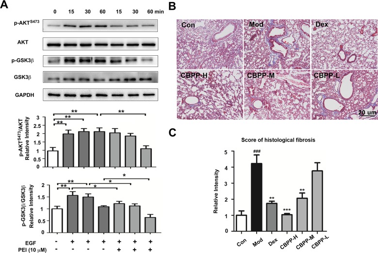
Figure 5.
PEI inhibited the EGFR signaling pathway, and CBPP alleviated pulmonary fibrosis. (A) BEAS-2B cells were pretreatment with PEI, then stimulated by 1 ng/ml EGF. The phosphorylation of AKTS473 and GSK3β was measured by Western blot, and the relative intensity data of P-AKTS473 and P-GSK3β to GAPDH are represented by the mean ± SD of three groups, *p < 0.05 and **p < 0.01. (B) Masson staining images (40-fold) of lung pathological sections and (C) statistics for the score of histological fibrosis, **p < 0.01 and ***p < 0.001 compared to the Mod. ### p < 0.001 compared to the Con (n = 6).

