Fig. 3.
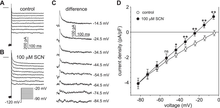
Determination of the voltage threshold for SCN− influx in retinal pigment epithelial (RPE) cells exposed to 100 µM external thiocyanate (SCN−). A and B: families of whole cell currents recorded in the same RPE cell in the absence (A) and presence (B) of 100 µM external SCN−. Currents were evoked by a series of voltage steps from a holding potential of −120 mV. The horizontal lines to the left of the current families represent the zero-current potential. The pipette and bath solutions contained 140 mM Cl− and 145.6 mM Cl−, respectively. C: family of SCN−-dependent currents obtained by taking the difference between the current traces in B from those in A. The value at the right of each current trace represents the amplitude of the voltage step, corrected for liquid junction potentials. D: current-voltage (I-V) relationships of peak current density obtained in the absence (○) and presence (●) of 100 µM external SCN−. Peak currents were measured 6–12 ms after the onset of the voltage steps. Symbols represent means and error bars represent SE; n = 5 cells from 3 C57BL/6J mice. *P < 0.05 compared with control; **P < 0.01 compared with control; ns, not significant, P > 0.05 (two-way ANOVA followed by Sidak’s multiple-comparisons test).

