Fig. 1.
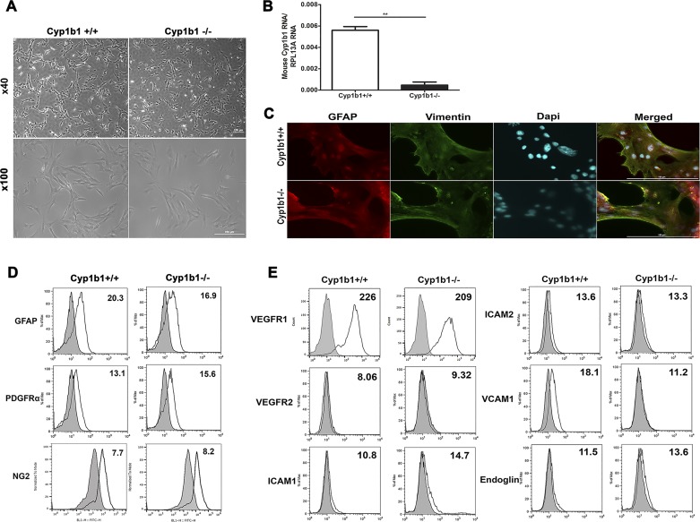
Isolation of mouse retinal astrocytes (ACs). Wild-type (Cyp1b1+/+) and cytochrome P450 1B1-deficient (Cyp1b1−/−) retinal ACs were isolated and cultured on gelatin-coated 60-mm dishes. A: cells were photographed in digital format at ×40 and ×100 magnification. Scale bars = 250 µm. Please note that there are no differences in retinal AC morphology regardless of Cyp1b1 status. B: expression of Cyp1b1 mRNA determined by quantitative PCR (**P < 0.01; n = 3). C: indirect immunofluorescence staining using an anti-vimentin and anti-paired-box protein Pax-2 (Pax2) antibody was performed as described in materials and methods. DAPI was used to stain cell nuclei. Greater than 98% of cells were positive for vimentin and Pax2 AC markers (n = 3; scale bars = 200 µm). D: Cyp1b1+/+ and Cyp1b1−/− ACs were examined for expression of other AC markers including glial fibrillary acidic protein (GFAP), PDGF receptor-α (PDGFRα), and neuroglia proteoglycan 2 (NG2) by flow cytometry. E: expression of VEGF receptor 1 (VEGFR1), VEGFR2, ICAM-1, ICAM-2, VCAM-1, and endoglin was also determined by flow cytometry. The representative mean fluorescence intensities are indicated in each histogram. Max, maximum; RpL13A, 60S ribosomal protein L13a.

