Fig. 8.
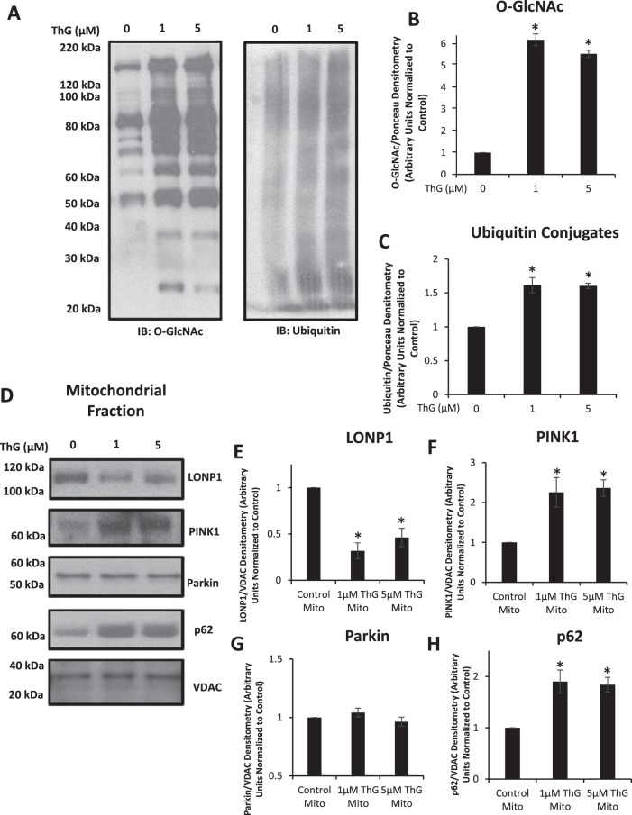
Effect of Thiamet-G (ThG) on ubiquitinated mitochondrial fractions. Mitochondrial fractions were isolated and assessed to measure changes in ubiquitin conjugates, or mitochondrial quality control proteins in response to ThG (1 and 3 µM) treatment for 6 h. The Millipore CTD 110.6 antibody was used in the mitochondrial O-linked β-N-acetylglucosamine (O-GlcNAc) blots shown here. A–C: Western blot analysis of O-GlcNAc (left) ubiquitin conjugates (right) and loading control staining are represented as well as quantification of the Western images normalized to the untreated control (B and C). D–H: Western blot analysis of Lon protease homolog 1 (LonP1), phosphatase and tensin homolog induced kinase 1 (PINK1), Parkin, and p62 are represented (D) as well as relative quantification of the Western images (E–H) normalized to the untreated control. Data are means ± SE; n = 3 replicates per sample. *P < 0.05, different from untreated control.

