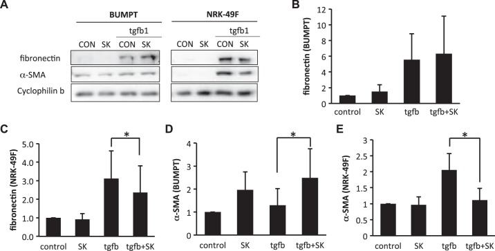
Fig. 8.
Divergent effects of shikonin (SK) on transforming growth factor (TGF)-β1-induced fibrotic changes in renal fibroblasts (NRK-49F) and proximal tubular cells (BUMPT) cells. BUMPT cells or NRK-49F cells were treated with or without 0.25 μM SK. Cells were exposed to 10 ng/ml TGF-β1 for 24 h to induce fibrosis. Whole cell lysates were collected and examined by immunoblot analysis. Protein expression levels from immunoblots were quantified by densitometry analysis and normalized with cyclophilin B expression. Data are expressed as means ± SD; n is the repetition number of parallel experiments. A: representative immunoblot images showing protein level changes of fibronectin and α-smooth muscle actin (α-SMA) with matched cyclophilin B as an internal marker for protein loading. CON, control. B: densitometry analysis of the fibronectin level in BUMPT cells. C: densitometry analysis of the fibronectin level in NRK-49F cells. n = 3, *P = 0.026. D: densitometry analysis of the α-SMA level in BUMPT cells. n = 3, *P = 0.029. E: densitometry analysis of the α-SMA level in NRK-49F cells. n = 3, *P = 0.028.

