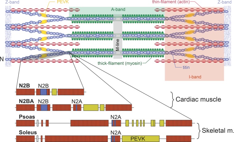
Fig. 3.
Schematic illustration of a sarcomere with Z band, actin, myosin, and titin filaments in their supposed arrangement. Titin runs from the middle of the sarcomere (M line) in either direction toward the Z band. Titin is thought to be firmly attached to myosin and then run freely across the I-band region of the sarcomere until ~50–100 nm away from the Z band, where it is thought to attach to actin and then enter the Z band bound to actin. Below the schematic sarcomere are illustrations of the structure of titin in cardiac muscle and in 2 selected skeletal muscles (psoas and soleus). Note that cardiac titin isoforms are much smaller than those of skeletal muscles and that skeletal muscles have different isoforms that are thought to reflect the passive requirements of the muscle. Specific segments (N2B, N2A, N2BA, and PEVK) are indicated. The brownish rectangles represent immunoglobulin domains. Adapted from Granzier and Labeit (16) with permission from Wolters Kluwer Health, Inc.

