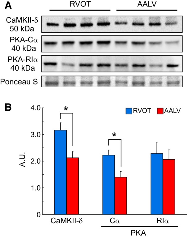
Fig. 10.

CaMKII-δ and PKA proteins are differentially expressed in the right ventricle (RV) and left ventricle (LV). A: representative Western blots of tissue extract from the RV outflow tract (RVOT) and the anterior apical LV (AALV). B: quantification of Western blotting results for CaMKII-δ, PKA-Cα, and PKA-RIα, showing that the expression levels of CaMKII-δ, PKA-Cα were significantly higher in the RVOT as compared with the AALV, whereas PKA-RIα exhibited a uniform regional distribution. Quantification of the enzymes’ regional expression levels was achieved by using the Ponceau S signals for normalization. A.U., arbitrary units. *P < 0.05, paired Student’s t-test; RVOT: n = 4; AALV: n = 4.
