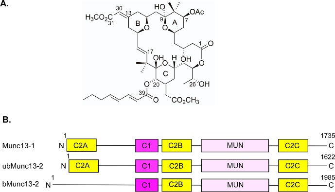
Figure 1.
Structures of bryostatin 1 and Munc13s. (A) Chemical structure of bryostatin 1. (B) Domain structure of Munc13-1, ubMunc13-2, and bMunc13-2. The C1 domain binds lipids and DAG/phorbol ester. C2 domains bind lipids and Ca2+. MUN is a self-folding domain consisting of two Munc13 homology domains. Constructs used during experiments contained a green fluorescent protein (GFP) tag at their C-termini.

