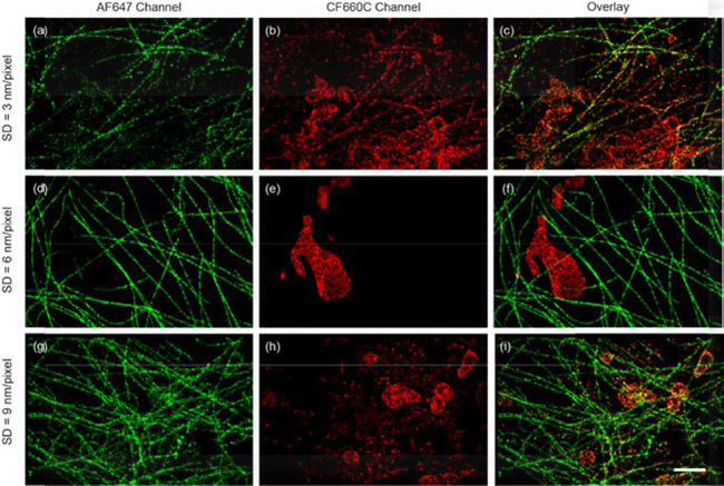
Fig. 3.
Two-color sSMLM images of AF647 labeled microtubule and CF660C labeled mitochondria in COS-7 cells at different SDs (a-c, 3 nm/pixel), (d-f, 6 nm/pixel) and (g-i, 9 nm/pixel) respectively. The AF647 (a, d and g) and CF660C (b, e, and h) channels and the overlay images (c, f and i) were collected with single-molecule SC of 683–689 nm and 692–698 nm respectively. (Scale bar = 2 μm)

