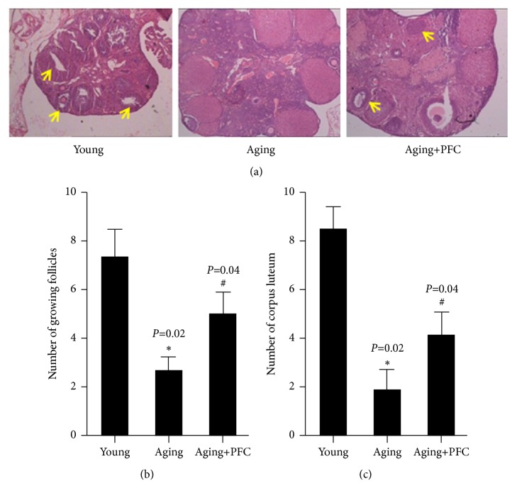
Figure 1.
PFC improves ovarian histology and increases follicles and corpus luteum in aging mice. (a) HE staining with ovarian tissues. The yellow arrows indicate the growing follicles. (b) Quantification of growing follicles in ovarian tissues. (c) Quantification of corpus luteum in ovarian tissues. For statistical significance in this figure, ∗P < 0.05 compared with the young control group; #P < 0.05 compared with the aging model group; Kruskal-Wallis H test.

