
Figure 6.
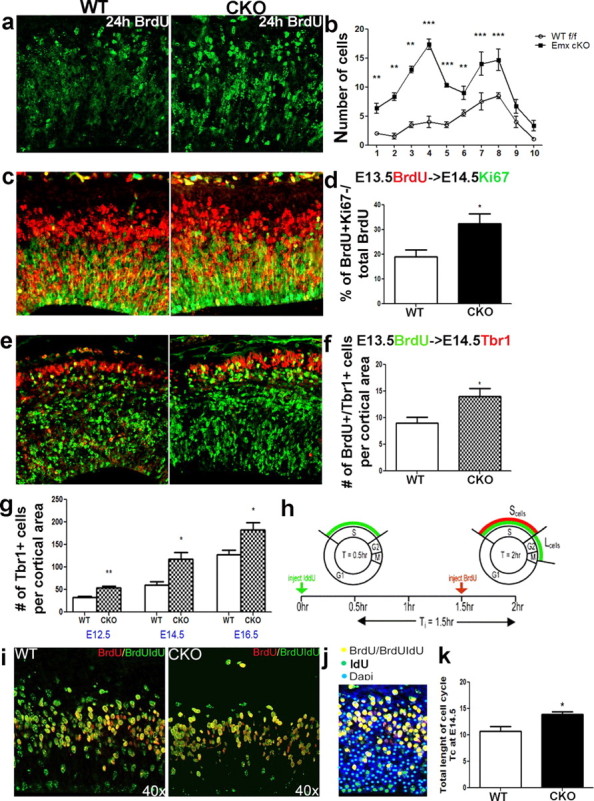
ERK2 CKO neural progenitors exhibit premature cell cycle exit and lengthening of the cell cycle during midneurogenesis. A single BrdU injection was administered at E13.5 and embryos were fixed 24 h later. Coronal sections were immunolabeled with BrdU (green). a, b, Cells that reenter the cell cycle show distinct puncta and are less intensely labeled, whereas cells that exited the cell cycle and become postmitotic are brightly labeled (a). Brightly labeled BrdU+ cells (at least 80% of cell covered) were counted across the cortical anlage (b). The cortical entity was divided into 10 vertical bins of equal thickness. The histogram shows a statistical analysis of the number of brightly labeled cells in each bin in CKO and WT cortex (a, b) (n = 3; p ≤ 0.001). c, d, Immunostaining with Ki67 (green) and BrdU (red) antibodies after 24 h BrdU pulse at E13.5. All cells that exited the cell cycle (BrdU+/Ki67−) were counted and their percentage of total BrdU+ cells evaluated 24 h postinjection was analyzed (d). e, f, To evaluate the fate of newly born cells, we colabeled with a postmitotic marker, Tbr1 (red) and BrdU (green), 24 h after injection (WT, n = 4; CKO, n = 5; p = 0.0383). g, The total number of Tbr1+ postmitotic neurons was also evaluated at E12.5 (p = 0.0034), E14.5 (p = 0.0007), and E16.5 (p = 0.0140). h, Cell cycle during midneurogenesis. Diagram adopted from Martynoga et al. (2005) showing a schematic of the double injection [iododeoxyuidine (IdU)/BrdU] paradigm. Pregnant dams were injected with IdU and BrdU 1.5 h apart and sacrificed 0.5 h later. i, Coronal sections were immunolabeled with IdU (green) and BrdU (red) at E14.5. k, Cells in the S phase by t = 2 h were labeled with both BrdU and IdU (S cells: yellow) (p < 0.05). j, Cells were counted in 100 μm-wide bins and are shown as colored dots.