Figure 2.
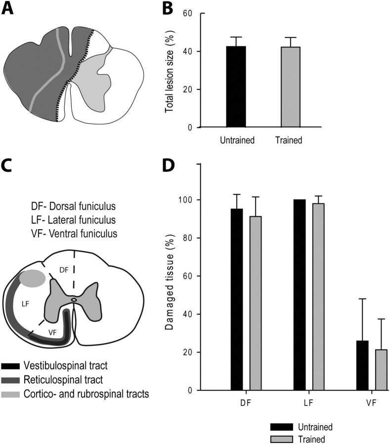
Comparison of the partial lesion size between groups. A, Schematic drawings of the largest and smallest hemisections reported in all cats (in gray, respectively bordered with black and white dotted lines). B, Comparison of the extent (%) of the total damaged area in the two groups. C, Schematic illustration of the subdivisions of the left hemicord into functional quadrants and of the descending pathways in the cat. The left side of the spinal cord was divided into three parts, as an attempt to delineate spinal white matter areas known a priori to contain descending fiber tracts that may subserve specific functions. The dorsal funiculus mainly contains the dorsal column tract that conveys sensory information from periphery to the contralateral somatosensory cortex. The lateral funiculus contains the descending cortico- and rubrospinal tracts and is mostly involved in voluntary and fine distal motor control. The ventral funiculus contains the reticulo- and vestibulospinal tracts, respectively, from the reticular formation and Deiter's nucleus, that are known to be involved in posture and locomotion as well as some direct corticospinal fibers. D, Comparison of the extent (%) of spinal white matter damage in specific functional funiculi in the two groups.

