Figure 1.

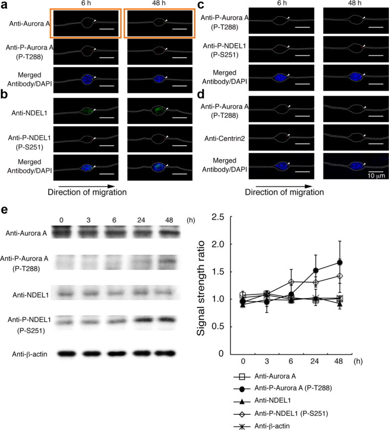
Aurora-A is expressed and phosphorylated NDEL1 in granular neurons. a, Examination of subcellular localization of Aurora-A (top), phospho-T288 Aurora-A (middle), and DAPI (bottom). Granular neurons displayed condensed expression (arrowhead) of Aurora-A (top) and phospho-T288 Aurora-A (middle). Time (in hours) after the start of culture is indicated above the panels. The white arrowheads indicate antibody signal. Phosphorylation of Aurora begins early after the start of migration. The white dotted lines indicate the outline of granular neurons. b, Examination of subcellular localization of NDEL1 (top), phospho-S251 NDEL1 (middle), and DAPI (bottom) is shown, and the white arrowheads indicate accumulation of these proteins. c, Comparison of phospho-T288 Aurora-A (top), phospho-S251 NDEL1 (middle), and DAPI (bottom). d, Comparison of phospho-T288 Aurora-A (top), Centrin-2 (middle), and DAPI (bottom). At early stage and later stage of neuronal migration, phosphorylated Aurora-A overlapped with Centrin-2, and the centrosomes were located at the front of the migrating nucleus. e, Western blotting analysis of each protein or phosphorylated protein. Either Aurora-A or NDEL1 displayed similar expression levels during culture, whereas phosphorylated Aurora-A or NDEL1 were increased during culture. Intensity was normalized to β-actin. Statistical analysis of three independent experiments is shown.
