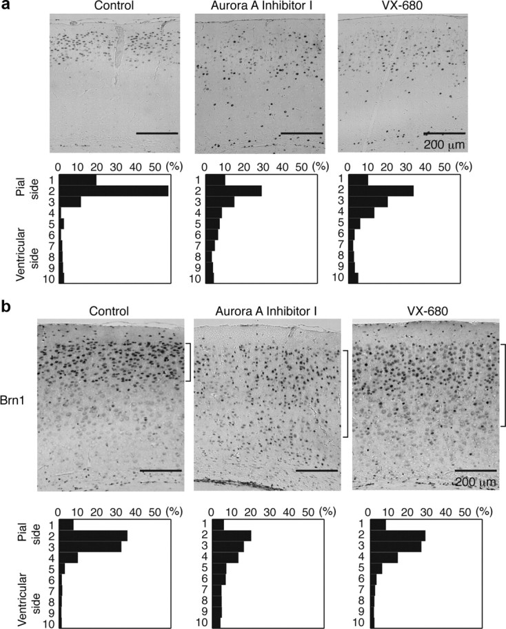
Figure 5.

Defective neuronal migration by intraperitoneal injection of Aurora-A inhibitors. a, BrdU birth-dating analysis revealed neuronal migration defects after intraperitoneal injection of Aurora-A inhibitors. Mice were injected with BrdU at E14.5 and killed at P21. Aurora-A inhibitors were injected twice at E15.5 and E17.5. Quantitative analysis was performed by measuring the distribution of BrdU-labeled cells in each of 10 bins that equally divided the cortex from the molecular layer (ML) to the subplate (SP) (bottom panels). The staining patterns are representative of 10 different experiments. Note the shift downward toward the ventricular surface after intraperitoneal injection of Aurora-A inhibitors. b, The distribution of Brn-1-positive cells is indicated at the right side of each panel. Brn-1-positive cells were more dispersed after intraperitoneal injection of Aurora-A inhibitors. Quantitative analysis was performed by measuring the distribution of Brn-1-positive cells in each of 10 bins that equally divided the cortex from ML to SP (bottom panels). The staining patterns are representative of 10 different experiments.
