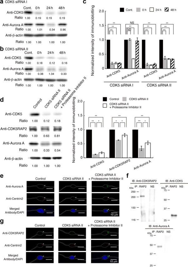
Figure 9.

CDK5 regulates centrosomal targeting of Aurora-A via CDK5RAP2. a, Granular neurons were transfected with siRNA I against CDK5, followed by incubation for 24 h to make cell aggregations. Cell aggregations of granular neurons were plated onto coated dishes (see Materials and Methods). Western blotting was performed at 0, 24, and 48 h after the start of culture. CDK5 was clearly reduced at the start of culture. Aurora-A was also reduced at 24 h after the start of culture, followed by a recovery 48 h after start of culture. Control indicates granular neurons before transfection. b, Granular neurons were transfected with siRNA II against CDK5. Western blotting was performed at 0, 24, and 48 h after start of culture. CDK5 and Aurora-A were reduced at the start of culture. Note: Aurora-A reduction continued 48 h after the start of culture. c, Relative intensity of the bands of Western blotting is displayed. Intensity was normalized with β-actin. Statistical examination was performed by unpaired Student's t test, with *p < 0.05 and **p < 0.01. d, Aurora-A reduction was prevented by administration of Proteasome Inhibitor II at a concentration of 0.5 μm. Proteasome Inhibitor II was applied at the time of siRNA transfection. Note: Proteasome Inhibitor II had no influence on the depletion of CDK5, whereas Proteasome Inhibitor II partially prevented degradation of Aurora-A. We also used other proteasome inhibitors. However, given the toxicity of these compounds to granular neurons, we were not able to continue the experiments in culture. Relative intensity of the bands of Western blotting is displayed at the bottom. Intensity was normalized with β-actin. Statistical examination was performed by unpaired Student's t test, with *p < 0.05 and **p < 0.01. e, Subcellular distribution of Aurora-A transfected with siRNA II in the presence of Proteasome Inhibitor II. Twenty-four hours after the start of culture, there was loss of centrosomal targeting and broader localization of Aurora-A. The white dotted lines indicate the outline of granular neurons. Centrin-2 was used as a centriole marker. f, Immunoprecipitation assay using an anti-CDK5RAP2 antibody. After coimmunoprecipitation, endogenous CDK5RAP2 was detected (right). CDK5 (middle) and Aurora-A (left) were also coprecipitated with CDK5RAP2. N.S. indicates nonimmune serum. g, Subcellular distribution of CDK5RAP2 24 h after the start of culture in the presence of Proteasome Inhibitor II. There was a loss of centrosomal targeting and broader localization of CDK5RAP2. The white dotted lines indicate the outline of granular neurons.
