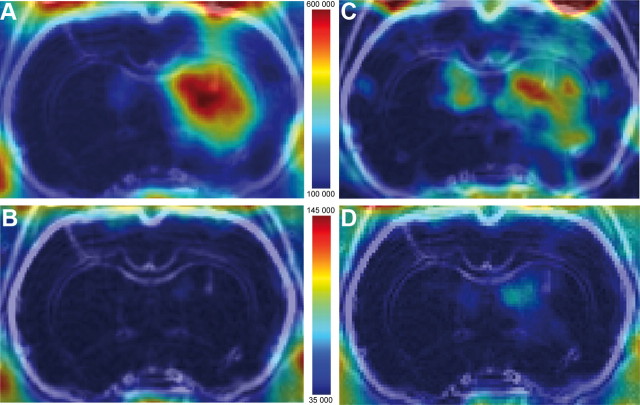
Figure 4.
Representative PET images of [18F]DPA-714 presaturation and displacement. Coronal rat brain views of [18F]DPA-714% SUV summed PET images (over 90 min), coregistered with the individual MRI under baseline (A), presaturation (B), and displacement conditions (C: %SUV summed image over first 15 min; D: %SUV summed image over last 75 min). Color-coded scale is in becquerel/cubic centimeter.

