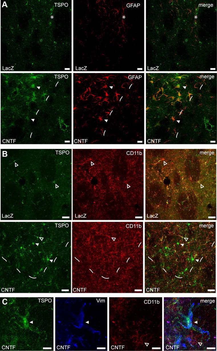
Figure 6.

TSPO is overexpressed by CNTF-activated astrocytes. A, In the striatum injected with lenti-LacZ, there is a low level of staining for GFAP (red) and TSPO (green). The needle track is indicated by a star. In the striatum injected with lenti-CNTF, TSPO staining is very intense and matches the area displaying GFAP-positive reactive astrocytes (area left to dashes). TSPO is expressed at the highest level in GFAP-positive astrocytes (white arrowhead). Scale bars, 20 μm. B, On the contrary, in the lenti-CNTF-injected striatum, there is only a limited colocalization of TSPO (green) with CD11b-labeled microglia (red, open arrowheads). Such colocalization is also observed in the control striatum injected with lenti-LacZ and gives rise to minimal PET signal. CD11b staining is not visibly enhanced in the area with reactive astrocytes (area above dashes). Scale bars, 20 μm. C, At higher magnification in the lenti-CNTF-injected striatum, vimentin-positive reactive astrocytes (blue, white arrowhead) express high level of TSPO (green), contrary to CD11b-labeled microglia (red, open arrowhead) that express undetectable levels of TSPO. Scale bars, 10 μm.
