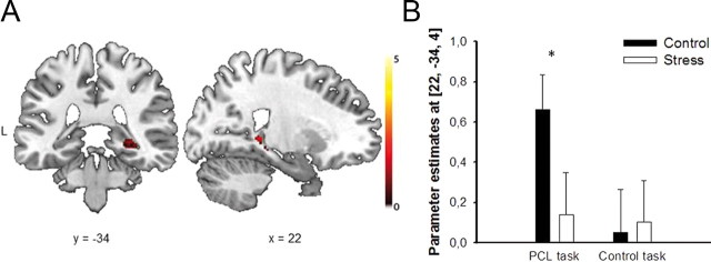
Figure 3.
Stress effect on hippocampal activity during classification learning. A, During the PCL task, the right hippocampus was significantly less activated in the stress group than in the control group (pcorr < 0.05, FWE corrected). Coronal and sagittal sections are shown, superimposed on a T1-template image. L, left. B, Parameter estimates of the peak voxel for the control and stress groups in the PCL and control task, respectively. Data represent mean ± SEM (*p < 0.05).

