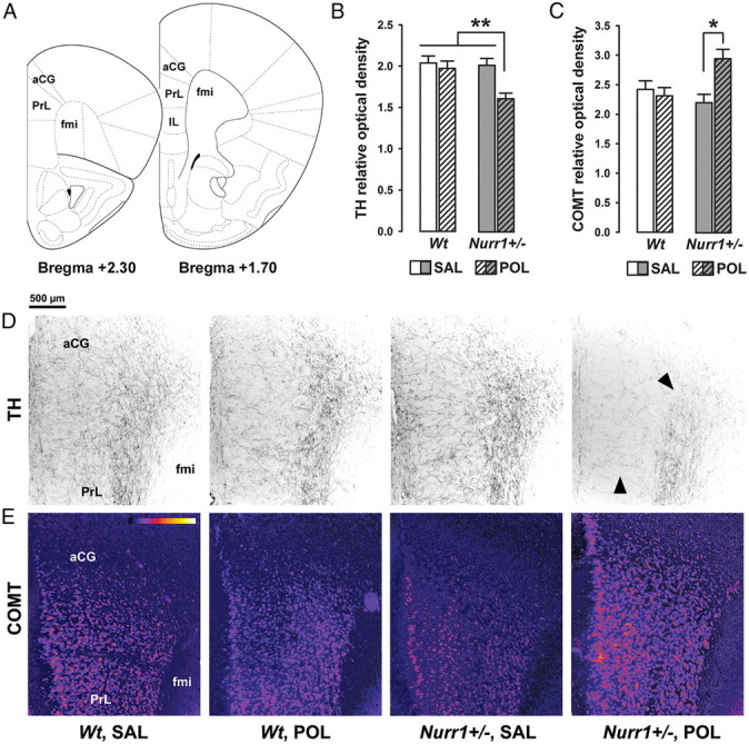
Figure 8.

Synergistic effects of genetic Nurr1 deficiency and prenatal immune activation on the prefrontal cortical dopamine system. A, Schematic coronal brain sections delineating the prefrontal cortical areas investigated with reference to bregma [adapted from The Mouse Brain in Stereotaxic Coordinates by Franklin and Paxinos (2008)]. Assessment of the relative optical densities of dopaminergic markers in the mPFC included measurements in the aCG, PrL, and IL cortices and was performed on coronal sections ranging from bregma +2.30 to +1.70 mm. B, Mean + SEM relative optical density of TH in the mPFC of adult wt and Nurr1+/− offspring prenatally treated with poly(I:C) (POL) (2 mg/kg, i.v.) or vehicle [saline (SAL)] solution on gestation day 17. **p < 0.01; N = 7 males in each experimental group. C, Mean + SEM relative optical density of COMT in the mPFC of SAL- or POL-exposed wt and Nurr1+/− offspring. *p < 0.05; N = 7 males in each experimental group. D, Coronal brain sections of representative SAL- or POL-exposed wt and Nurr1+/− offspring stained with anti-TH antibody. The sections were taken at the level of the mPFC highlighting TH-positive fibers in the aCG and PrL cortices. Note the marked decrease (indicated by the arrowheads) of TH-positive fibers emerging selectively in POL-exposed Nurr1+/− offspring. E, Coronal brain sections of representative SAL- or POL-exposed wt and Nurr1+/− offspring stained with anti-COMT antibody. The sections were taken at the level of the mPFC highlighting COMT protein expression in the aCG and PrL cortices. The sections were color-coded for the purpose of facilitating the visualization of differential COMT protein expression; strongest staining intensities are shown in yellow, while the background is represented in dark purple (bar inset). fmi, Forceps minor corpus callosum.
