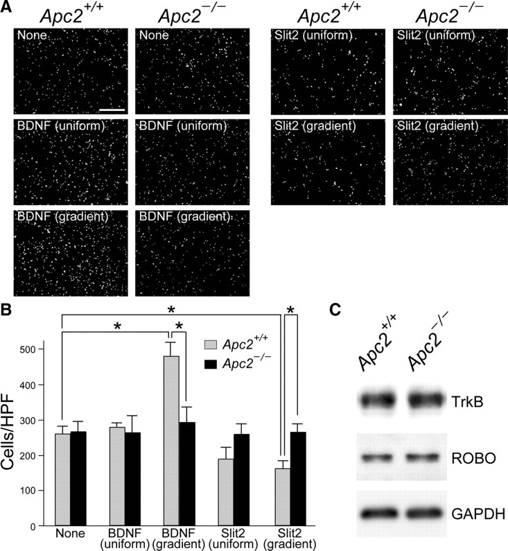
Figure 13.
Impaired response of Apc2-deficient CGCs to a BDNF or Slit2 gradient. Purified wild-type and Apc2-deficient CGCs were grown for 16 h in Boyden chambers with or without 30 ng/ml BDNF or 50 ng/ml Slit2. The factors were added to both compartments (uniform) or only to the lower compartment (gradient). CGCs that migrated through the porous membrane into the lower chamber were stained with DAPI and quantified. A, Representative images of migrated CGCs under distinct conditions. Scale bars, 200 μm. B, Quantification was performed by counting migrated cells per high-powered field (cells/HPF ± SEM) in 10 fields from replicate wells. Gray bars, wild-type CGCs; black bars, Apc2-deficient CGCs. The asterisk indicates a significant difference between the two values by Student's t test (*p < 0.05). C, Western blot analyses of TrkB, Robo, and GAPDH in extracts of purified CGCs.

