
Figure 2.
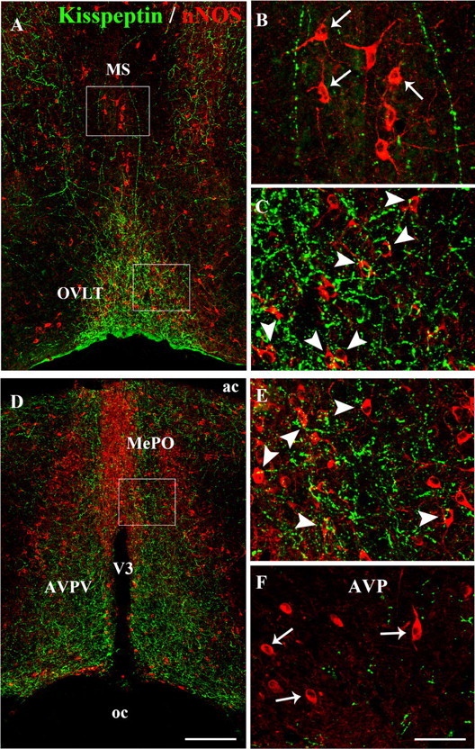
Kisspeptin neurons project onto nNOS neurons in the preoptic region. A, Low-magnification photomontage of kisspeptin (green) and nNOS (red) immunofluorescence in the hypothalamic preoptic region at the level of the OVLT and the MS. B, C, High-magnification images of the boxed areas in A, showing kisspeptin-immunoreactive fibers (green) and nNOS-immunoreactive cell bodies (red) in the MS (B) and the OVLT (C). Note that kisspeptin fibers are abundantly apposed to nNOS neurons in the OVLT (C, arrowheads), but not in the MS (B, arrows). D, Low-magnification photomontage of kisspeptin (green) and nNOS (red) immunofluorescence in the hypothalamic preoptic region at the level of the MePO and the anteroventral periventricular nucleus (AVPV). E, F, High-magnification images of the boxed area in D, showing kisspeptin-immunoreactive fibers (green) and nNOS-immunoreactive cell bodies (red) in the MePO (E) and the AVP (F). Note that abundant kisspeptin fibers are apposed to nNOS neurons in the MePO (E, arrowheads), but not in the AVP (F, arrows). V3, Third ventricle; ac, anterior commissure; oc, optic chiasm. Scale bars: A, D, 200 μm; B, C, E, F, 50 μm.