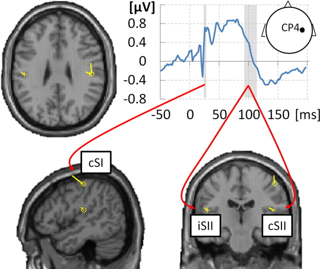
Figure 1.
Localizer ERP and resulting dipoles. Somatosensory-evoked potential at electrode CP4 (grand-averaged localizer data) is shown in top right plot. Individual N20 component latencies were used to fit a single dipole in cSI [average MNI location (44, −21, 55), SD (5, 12, 8)]. Adding to this dipole, a symmetrical dipole pair in cSII/iSII [average MNI location (±50, −18, 21), SD (10, 7, 13)] was fitted to the 90–115 ms time window. Resulting dipoles are shown, plotted on the T1 single-subject template (MNI), for a subject with the smallest mean Euclidean distance of the three dipoles from average locations.

