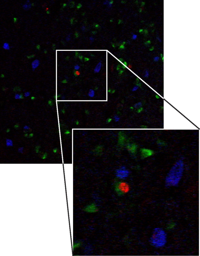
Figure 9.

Four-year-old adult-born neurons in HVC do not express interneuron markers. A mixture of antibodies against the calcium-binding proteins calbindin, calretinin, and parvalbumin stains many interneurons in HVC (blue) but there is no colocalization between the interneuron markers and BrdU (red) in birds that had received BrdU injections at 90 d of age and were killed 4 years later. HVC-RA neurons are labeled with CTB retrograde tracer injected into RA before killing (green). The enlarged panel shows the BrdU-positive nucleus of a retrogradely labeled HVC-RA neuron surrounded by three interneurons positive for the mix of calcium binding proteins.
