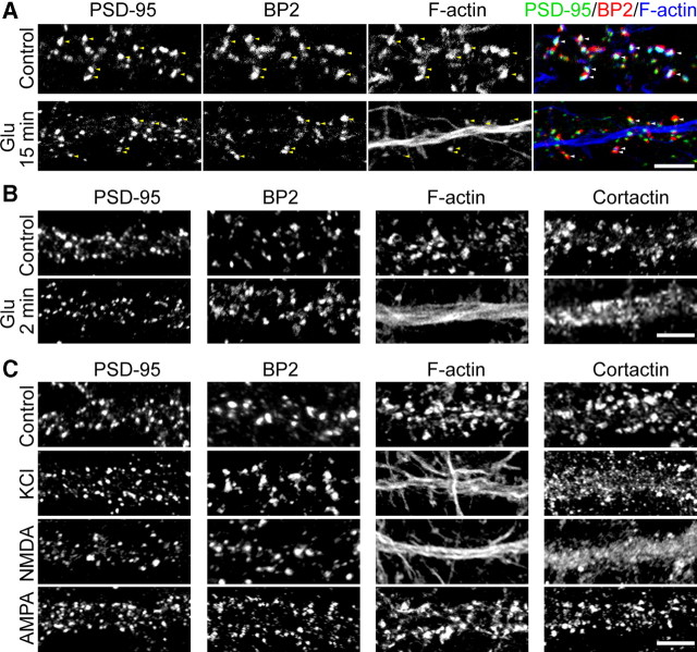
Figure 6.
CTTNBP2 stably resides at dendritic spines after glutamate stimulation. At 21∼24 DIV, cultured rat hippocampal neurons were stimulated with glutamate (50 μm), NMDA (100 μm), AMPA (100 μm), and KCl (75 μm) for 15 min (A), 15 min (C), or 2 min plus 8 min of recovery (B) in normal growth medium. A, Triple staining with PSD-95 antibodies, CTTNBP2 (BP2) antibodies, and phalloidin. Yellow arrowheads in the individual panels and white arrowheads in the merged panels point to the puncta containing overlapping PSD-95 and CTTNBP2. B, C, Distributions of PSD-95, CTTNBP2, F-actin, and cortactin along dendrites with or without stimulation. Due to technical limitations, we were unable to visualize PSD-95, CTTNBP2, cortactin, and F-actin in the same neurons. Images shown in B and C were not captured from the same cells. Scale bars, 5 μm.

