Figure 1.
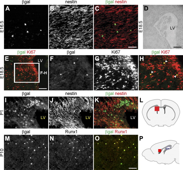
Runx1 is expressed in a restricted group of cells in the developing forebrain. A–H, Coronal sections through the telencephalon of either E16.5 (A–C) or E18.5 (D–H) Runx1LacZ/+ embryos were subjected to double-labeling immunofluorescence analysis of β-gal and nestin expression (A–C), determination of β-gal activity using X-gal staining (D), or double-labeling immunofluorescence analysis of β-gal and Ki67 expression (E–H). E, Box indicates area shown at higher magnification in F–H. H, Arrowheads indicate cells coexpressing β-gal and Ki67. I–K, Coronal sections through the telencephalon of P1 Runx1LacZ/+ pups were subjected to double-labeling immunofluorescence analysis of β-gal and nestin expression. β-gal+ cells do not express nestin but are located adjacent to nestin+ cells. L, Schematic of the forebrain in the coronal plane indicating the location (red square) of the area examined in panels E–K. M–O, Sagittal sections of P10 Runx1LacZ/+ brains were subjected to double-labeling analysis of β-gal and Runx1 expression. All β-gal+ cells express the Runx1 protein, indicating that β-gal expression faithfully recapitulates Runx1 expressed in the postnatal forebrain. P, Schematic of the forebrain in the sagittal plane demonstrating the location (red square) of the area examined in M–O. LV, lateral ventricle. Scale bars: (in C, O) A–C, F–K, M–O, 50 μm; E, 100 μm; D, 250 μm.

