
Figure 1.
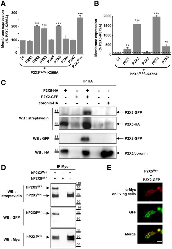
P2X5 subunits interact with P2X2 subunits at the plasma membrane. A, Rescue of cell surface expression of the P2X2–K366A trafficking deficient mutant by other subunits. P2X2–K366A carrying a Flag tag in the extracellular loop was expressed alone or in combination with each of the seven wild-type P2X subunits. Membrane expression was measured using a chemiluminescent assay. Cell surface expression of P2X2–K366A is rescued upon coexpression with P2X2, P2X3, P2X4, and P2X5 subunits. B, Membrane detection of the P2X5–K372A mutant carrying an extracellular Flag tag is increased when coexpressed with P2X1, P2X2, and P2X4 and P2X5 subunits. Note that because the surface expression of P2X5–K372 mutant is very low compared withP2X2–K366A, the changes appear much higher. Results are shown as mean ± SEM of at least three independent experiments. **p < 0.01, ***p < 0.005, Student's t test. C, P2X5 subunits interact with P2X2 at the plasma membrane. P2X5 carrying an extracellular HA tag was expressed alone or in combination with P2X2-GFP. Immunoprecipitation was performed after labeling living cells with sulfo-NHS-LC-biotin. Top, Biotinylated protein fraction after HA immunoprecipitation detected with streptavidin-HRP. Middle and bottom, Detection of coimmunoprecipitated GFP and HA tagged-proteins, respectively. Coronin was used as a control for cell permeabilization. D, Human P2X2 and P2X5 subunits interact at the plasma membrane. Experiments were carried as described above, except that human P2X2-Myc and human P2X5-GFP cDNAs were used. E, Immunodetection of P2X5 in living cells. HEK cells were transfected with P2X5Myc subunits carrying an extracellular Myc tag and P2X2-GFP. Immunostaining of P2X5Myc subunits (red) was performed by incubation of Myc antibodies on living cells; P2X2 subunits (green) were revealed through GFP fluorescence. Scale bar, 10 μm.