
Figure 2.
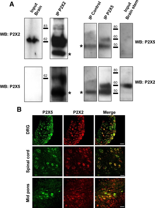
P2X5 and P2X2 subunits interact in neuronal tissues. A, Physical interactions of P2X2 and P2X5 subunits in brain tissues. P2X2 or P2X5 subunits were immunoprecipitated from mouse brain membrane and different subunits detected with specific antibodies. Left panel represents immunoprecipitation from total brain membrane proteins, right panel from brainstem. Note that P2X5 is not detectable from the total protein extract. Blots are representative of three independent experiments. Asterisks indicate the antibody heavy chains. B, Coimmunolocalization of P2X2 and P2X5 subunits in peripheral and central neurons. Representative images of immunohistochemistry performed on slices from DRG, spinal cord, or mid pons. P2X2 and P2X5 are stained in red and green, respectively. Note that in each structure only a subset of neurons coexpresses both subunits. Scale bar, 40 μm.