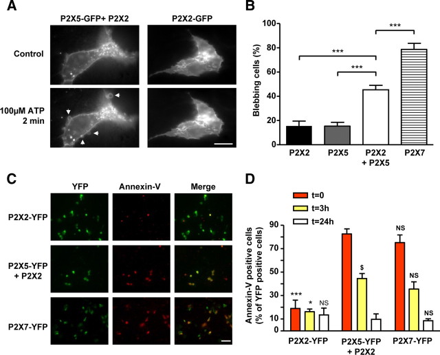
Figure 9.
Activation of P2X2/5 receptors mediates membrane blebbing and pseudo-apoptosis. A, B, P2X2/5 activation triggers membrane blebbing. A, Representative fluorescence images of HEK cells expressing P2X2-GFP or P2X2 + P2X5-GFP 2 min after 100 μm ATP stimulation. Arrows indicate ATP induced blebs. Scale bar, 10 μm. B, Quantitative analysis of the percentage of blebbing cells expressing P2X2-GFP, P2X5-GFP, P2X2/5-GFP, or P2X7-GFP after stimulation with 100 μm ATP (2 mm for P2X7) for 2 min. Results were normalized to the number of GFP-positive cells. Data are mean ± SEM of N = 3 independent experiments. ***p < 0.005, one-way ANOVA, followed by Bonferroni's multiple-comparison test. C, D, P2X2/5 receptor activation induces transient annexin-V exposure. C, HEK cells transfected with P2X2-YFP, P2X2+P2X5-YFP, or P2X7-YFP were stimulated with 100 μm ATP (500 μm for P2X7-YFP) and annexin-V staining was performed 5 min after the end of the stimulation. Scale bar, 50 μm. D, Analysis of annexin-V staining at 0, 3, or 24 h after ATP stimulation. Experiment was performed as in C, except that annexin-V staining was performed at the time indicated after ATP stimulation. In all panels, results were normalized to the number of YFP-positive cells. Data are mean ± SEM, n > 30 cells, N = 3 experiments. *p < 0.05, ***p < 0.005; N.S., not significant by comparison with P2X2/5 transfected cells at the same time point. $p < 0.05, comparison between P2X2/5-expressing cells at 0 and 3 h. One-way ANOVA, followed by Bonferroni's multiple-comparison test.

