Figure 1.
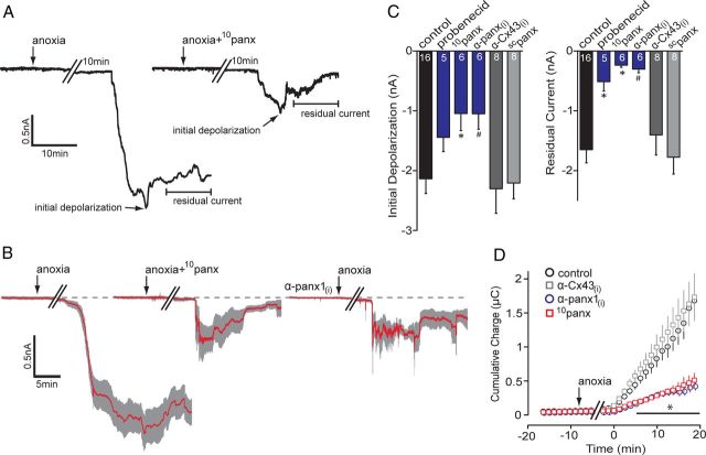
Block of pannexin-1 attenuates anoxia-induced inward currents. A, Representative whole-cell recordings from CA1 pyramidal neurons in hippocampal slices exposed to anoxia (onset indicated by arrows) with and without the presence of the Panx1 blocker 10panx (100 μm; right trace). Initial depolarization and residual currents are indicated (arrows) and were significantly attenuated by 10panx. B, Mean (red line) ± SEM (gray region) of 6–16 neurons (see C) of the AD in response to different Panx1 antagonists. Both 10panx (middle) and the intracellularly applied anti-Panx1 antibody, α-panx1(i) (0.25 ng/μl; right), reduced the AD-associated inward current, with promotion of recovery toward the baseline (dashed line). C, Quantitative analyses of the initial depolarization and residual current in the presence of Panx1 antagonists; the number of cells in each experiment are indicated in the bars. D, Cumulative net charge transfer during the anoxic inward current determined as area under the curve. Bath application of 10panx and α-panx1(i) both significantly attenuated the net charge compared with anoxia alone and their respective negative controls of bath-applied scrambled 10panx (scpanx, 100 μm) or intracellular anti-connexin 43, α-Cx43(i) (0.3 ng/μl; an α-panx1(i) control). *p < 0.05, significance compared with control; #p < 0.05, significance compared with α-Cx43(i). Error bars in B–D are SEM.

