
Figure 6.
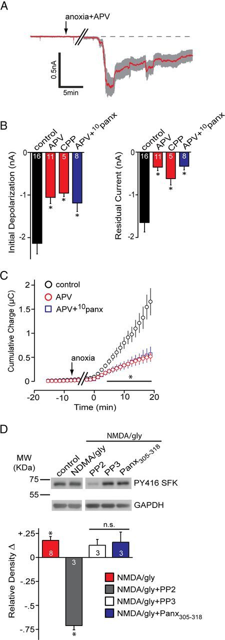
The contribution of NMDARs to Panx1 activation during the anoxic depolarization. A, Mean ± SEM trace of the anoxic depolarization during bath application of the NMDAR antagonist d-APV (50 μm). Blocking NMDARs promoted recovery toward the baseline. B, Quantitative summary of the anoxic depolarization with NMDAR block by d-APV or R-CPP (5 μm), as well as concomitant block of NMDAR and Panx1 (d-APV+10panx). Control data are the same as those presented in Figure 1. C, Net cumulative charge transfer across the membrane with NMDAR block by d-APV or R-CPP. D, Top, Src family kinase activation in the presence of bath-applied NMDA/glycine (gly) assayed by Western blot analysis of phospho-Y416 levels. Bottom, Quantitative analysis of the relative (vs control) density of phospho-Src Y416. Band densities were normalized to GAPDH and expressed relative to control, which was set at zero for each gel. Application of NMDA/glycine was significantly different than control (one-way Student's t test vs a mean of 0). PP2 was significantly different (p < 0.05) from NMDA/gly. All bands in the blot are from the same gel. Error bars are SEM.