
Figure 5.
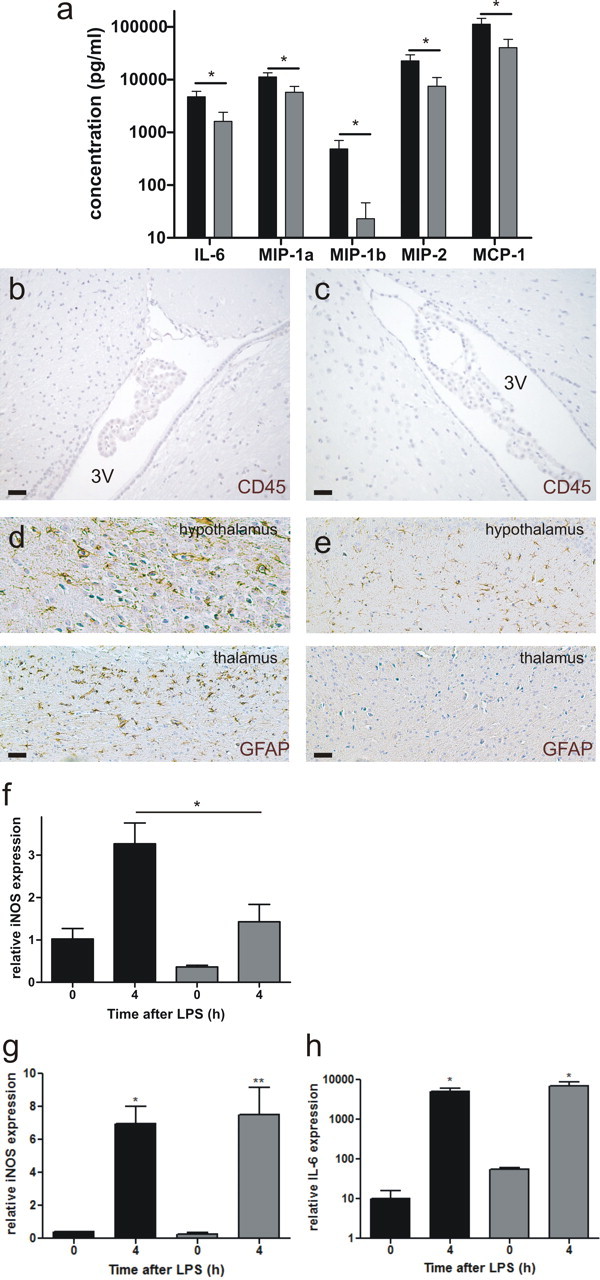
MMP8 depletion reduces the effect of peripheral inflammation on the CNS. a, Cytokine and chemokine profile in CSF isolated from MMP8+/+ mice (■, n = 8) and MMP8−/− mice ( , n = 8) 8 h after LPS challenge. Data are expressed on a log10 scale. Cytokines and chemokines were determined by BioPlex assay. Levels in unstimulated mice were negligible. Absence of white blood cell influx in the brain tissue surrounding the third ventricle (3V) in MMP8+/+ (b) and MMP8−/− (c) mice 8 h after endotoxemia induction, determined by CD45 immunohistochemistry (brown). Scale bar, 20 μm. GFAP-positive cells (brown) in the hypothalamus (d) and thalamus (e) of LPS-stimulated mice, 8 h after stimulus. Scale bar, 20 μm. f, Relative iNOS expression in the brain, before and 4 h after LPS injection in MMP8+/+ mice (■, n = 5) and MMP8−/− mice (, n = 5). iNOS (g) and IL-6 (h) mRNA levels in the CP before and 4 h after LPS challenge in MMP8+/+ (■) and MMP8−/− () mice (0 h, n = 3; 4 h, n = 5).