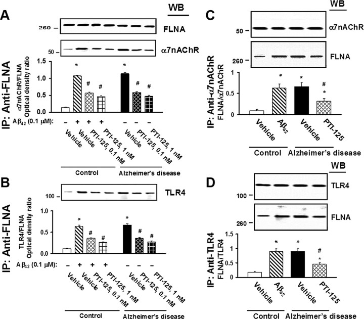
Figure 2.
A–D, Incubation for 1 h with 1 nm PTI-125 reduced FLNA–α7nAChR/TLR4 associations shown by Western blot detection of α7nAChR (A) or TLR4 (B) in anti-FLNA immunoprecipitates from AD and Aβ42-treated control FCX slices or by Western detection of FLNA in anti-α7nAChR (C) or anti-TLR4 (D) immunoprecipitates. Western blots (inset) were analyzed by densitometric quantitation. n = 11. *p < 0.01 vs vehicle-treated control; #p < 0.01 vs Aβ42-treated control or vehicle-treated AD.

